 |
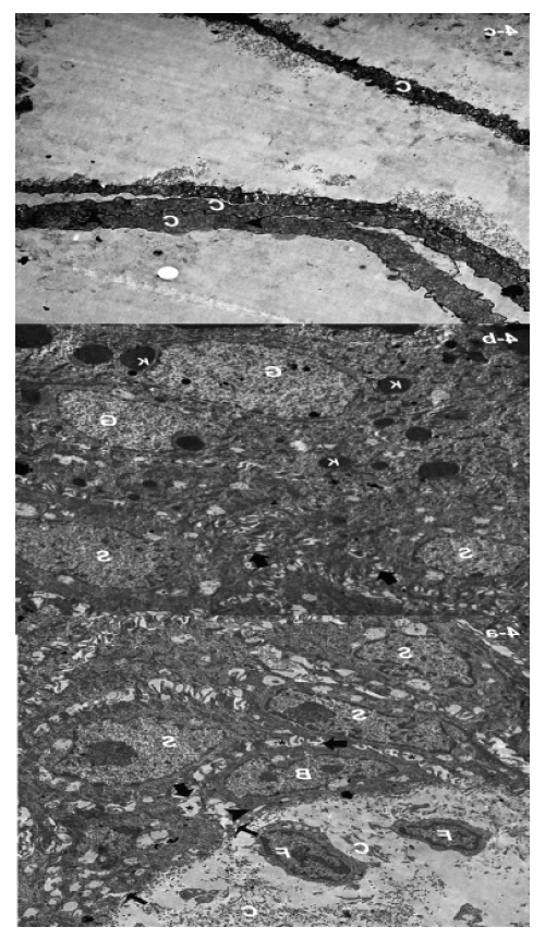
| Figure 4: Electron micrograph of the skin a ten day old rat showing: (a) Too little collagen fibres (C), fibroblast (F), cells of stratum spinosum (S) and cells of stratum basale (B). The basal cells are attached to each other’s by desmosomes (thick arrows) and to the basal lamina (thin arrows) by hemidesmosomes (arrowhead). Note narrow intercellular spaces (*) inbetween the stratum basale cells (X 4000). (b) Cells of stratum spinosum (S) with narrow intercellular spaces (*) in-betweens. The cells are attached to each other’s by desmosomes (arrows). A stratum granulosum cells (G) contains keratohyalin granules (K) (x 4000). (c) The stratum corneum cells (C) filled with electron-dense materials (arrowheads). |