|
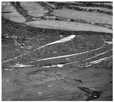
| Figure 6: Electron micrograph of rats’ skin showing: (a ) The stratum corneum cells (C) filled with electron-dense materials (arrowheads) of a two month rat’s skin. Note the keratohyalin granules (arrows) (4000X). (b) A well-developed collagen fibres (C) of a two month rat’s skin (5000X). (c) Collagen fibres (C) and elastic fibres (e) of a six month rat’s skin (3000X). |