 |
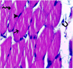
| Figure 2: Photomicrograph of a section in the skeletal muscle of a control rat (group I) showing: Normal histological architecture of the skeletal muscle fibers which appear transversely cut (arrow) with multiple, long and peripheral nuclei (arrowhead). Note the connective tissue endomysium separating the muscle fibers (wavy arrow) and the connective tissue perimysium separating the muscle bundles (curved arrow). Hx&E 400x. |