|
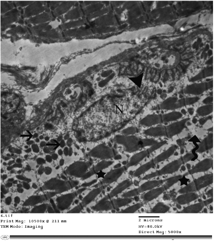
| Figure 15: Electron micrograph of a skeletal muscle of a rat of group II showing loss of regular arrangement of the myofibrils, fragmentation of the myofibrils (wavy arrow) with increased inter myofibrillar space (star), electrondense mitochondria (arrows) and irregular outline of the nuclear membrane (arrowhead). X5000. |