|
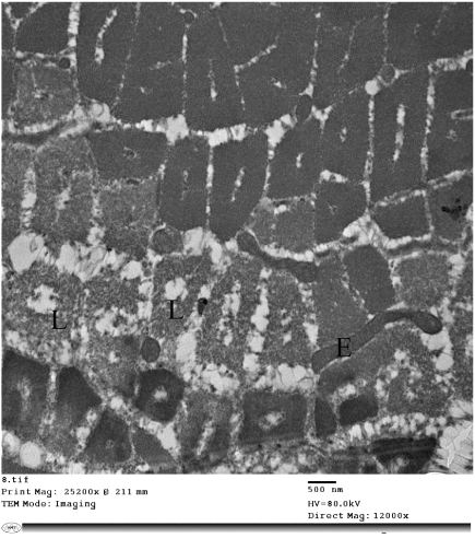
| Figure 18: Electron micrograph of a skeletal muscle of an ischemic rat showing transversely oriented myofibrils exhibiting cytoplasmic lysis (L) with complete loss the normal architecture. Note the presence of intermyofibrillar exudate (E). (X12000). |