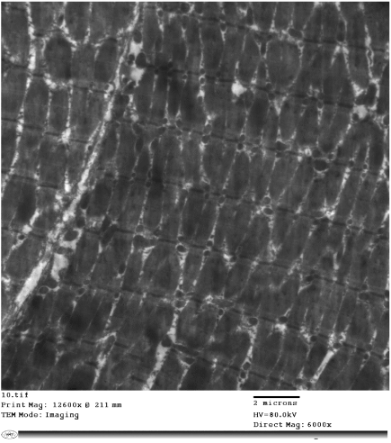
Figure 22:
Electron micrograph of a skeletal muscle of a rat of group IV showing apparently normal arrangement of most of the myofibrils. X6000.