|
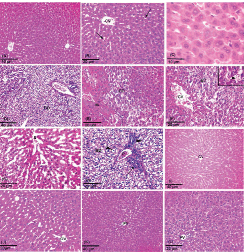
| Figure 1: (A-C) Photomicrographs of control rat liver from subgroup (i) (A) Showing normal hepatic architecture (H&E, Mic. Mag. 200X). (B) Showing polyhedral hepatocytes with eosinophilic cytoplasm arranged in strands around the central vein (CV), presinusoidal spaces (→) appears between the cords of hepatocytes (H&E, Mic. Mag. 400X). (C) Showing polyhedral hepatocytes with rounded vesicular nuclei and prominent nucleoli (→). Notice Kupffer cell (>) (H&E, Mic. Mag. 1000X). (D-H) Photomicrographs of liver sections of acetaminophen -treated group exhibit severe damage in the hepatic architecture. Most of hepatocytes fused together forming eosinophilic syncytial (M) masses. Dilatation of central vein (CV), blood sinusoid (S) and portal venule (P) are seen ballooning degeneration of hepatocytes (BD) is seen with vacuolated cytoplasm and indistinct cell boundaries. Notice mononuclear cellular infiltration (→) and apoptotic hepatocytes (>) around the portal venule (H&E, Mic. Mag. D, 200× and E-H, 400X).The inset showing vacuolated hepatocytes with lost nucleus and discontinuous cell boundaries (H&E, Mic. Mag. 1000×). (I-J) Photomicrographs of a section of the liver of acetaminophen and N-acetylcysteine treated group showing normal architecture of hepatic lobule and apparently normal looking hepatocytes with central vesicular nuclei around the central vein (CV). (K-L) Photomicrograph of liver section in the acetaminophen and curcumin treated group showing normal architecture of hepatocytes, portal venule (PV), and sinusoids (H&E, Mic. Mag. I&K, 200× and J&L, 400X). |