|
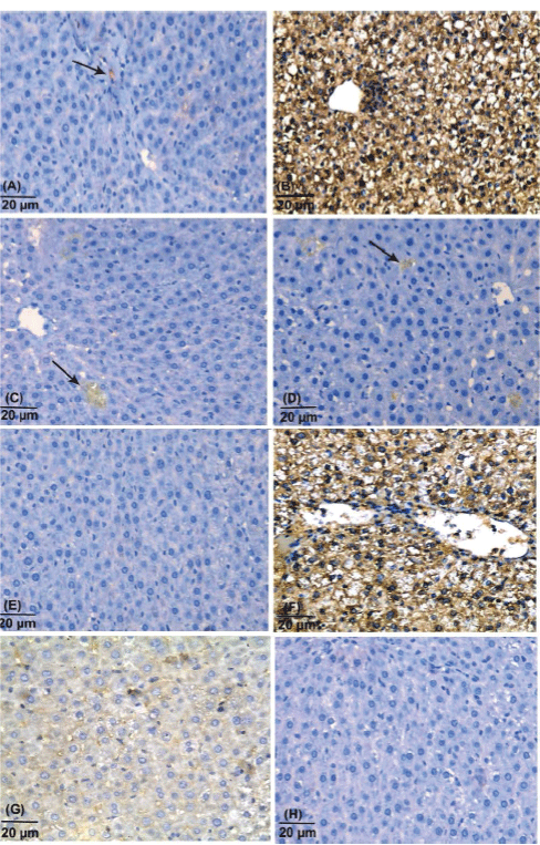
| Figure 2: (A-D) p53 immunostaining. (A) A section of the liver of control group from subgroup (i) showing faint and few cytoplasmic reaction for p53 (→). (B) A photomicrograph of a section of the liver of acetaminophen treated group showing strong p53 immunoreactivity in almost all hepatocytes seen. (C) A section of the liver of acetaminophen and N-acetylcysteine treated group showing weak p53immunoreativity in some hepatocytes (→). (D) A section of the liver of acetaminophen and curcumin treated group showing weak p53 immunoreactivity in some hepatocytes (→) ( p53 immunostaining & hematoxylin counterstain, Mic. Mag. 400X). (E-H) iNOS immunostaining. (E) A section of the liver of control group from subgroup (i) showing barely detected iNOS proteins in cytoplasm of hepatocytes. (F) A photomicrograph of a section of the liver of acetaminophen treated group showing strong expression of iNOS proteins in almost all hepatocytes. (G) A section of the liver of acetaminophen and N-acetylcysteine treated group showing weak expression of iNOS in hepatocytes. (H) A section of the liver of acetaminophen and curcumin treated group showing barely detected iNOS proteins in cytoplasm of hepatocytes (iNOS immunostaining & hematoxylin counterstain, Mic. Mag. 400X). |