 |
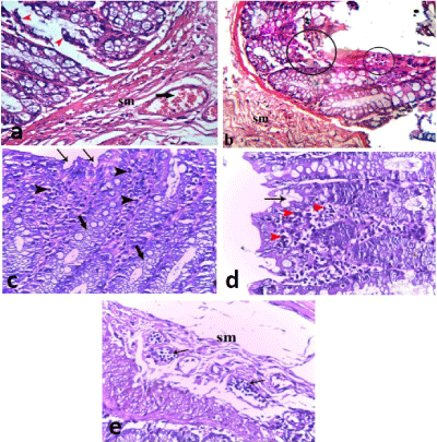
| Figure 2: Photomicrographs of acetic acid colitis group showing: (a, b) sloughed epithelial cells (arrowheads), pyknotic nuclei (encircled in b) and widened submucosa (sm) with capillary congestion (thick arrow). (c) Irregular intestinal surface (arrows) and dilated intestinal crypts lined by vacuolated cells (thick arrows). The mucosa shows inflammatory cellular infiltration (arrow heads). (d) Shallow intestinal crypts (arrow) and mucosal inflammatory cellular infiltration (arrow heads). (e) Dilated congested lymphatic vessels (arrows) within widened submucosa (sm). (H & E, 400X). |