 |
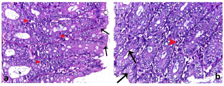
| Figure 4a, 4b: Photomicrographs of colon sections of NAC and ginger treated group showing the mucosa lined with normal intestinal epithelial cells (arrows) with intact mucosal surface. Few inflammatory cells (arrow heads) are dispersed within the mucosa. (H & E, 400X) |