|
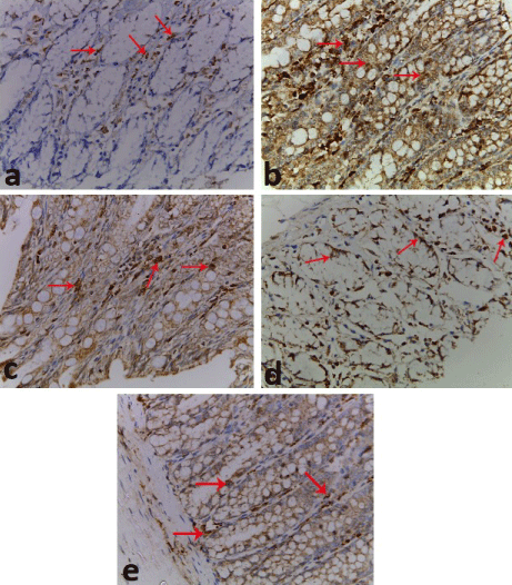
| Figure 7: Photomicrographs of sections in the colon showing: (a) control group with small number of i.NOS stained cells dispersed throughout the mucosa and within the fibrous tissue of the lamina propria with diffuse cytoplasmic staining (arrows). (b) Acetic acid colitis group with large number of i.NOS stained cells (arrows). (c,d) NAC treated and ginger treated groups respectively, with marked decrease of immunopositive cells (arrows), more in ginger group. (e) NAC and ginger treated group with mild number of immunopsitive cells (arrows). (i.NOS immunostaining, 400X). |