 |
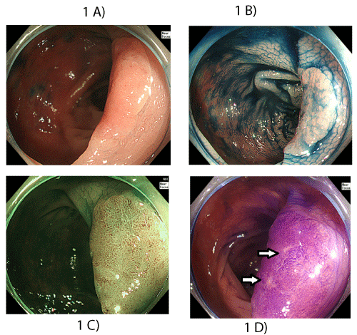
| Figure 1: A. Conventional endoscopy revealed a reddish, elevated lesion approximately 20 mm in diameter. B. Chromoendoscopic view with indigocarmine solution revealed a well-demarcated lesion margin. C. The surface of the lesion was observed by ME with NBI to be glandular structures of relatively uniform size without abnormal microvessels. D. Chromoendoscopic view of the surface with crystal violet staining revealed a type II pit pattern and partially a type Vi pit pattern in the center of the lesion (white arrow) (Kudo’s classification). |