magnifications 400X.
|
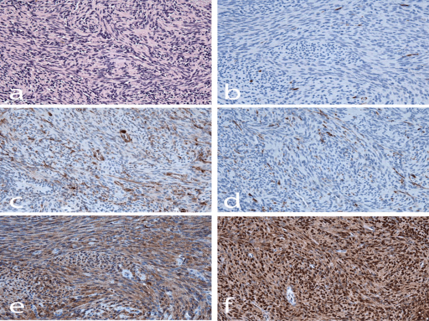
| Figure 2: (a) Higher magnification H&E stain of tumor demonstrates elongate spindle cells tightly arranged in fascicles and whorls in an Antoni A-type pattern. (b) CD117 (c-Kit) IHC is negative except for scattered infiltrating mast cells. (c) Smooth muscle actin highlights rich intratumoral capillary network and thin supporting stromal branches; tumor cells are negative. (d) Desmin is negative except for some supporting stromal cells. (e) Neuron-specific enolase uniformly labels tumor cells predominantly within the cytoplasm. (f) S-100 uniformly labels tumor nuclei intensely and cytoplasm moderately. All magnifications 400X. |