|
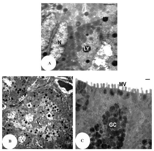
| Figure 4: TEM micrograph of the oesophagus mucosal cells of Natrix tessellata, note, (a) nucleus (N) and lysosomes(LY). (Scale bar, 2 μm). (b) the basal lamina (BL), mitochondria (M) and vacuoles (V). (Scale bar, 500 nm). (c) microvilli (MV) and the goblet cells (GC). (Scale bar, 500 nm). |