 |
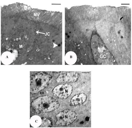
| Figure 11: TEM micrograph of the intestine mucosal cells of Scincus scincus, showing (a) microvilli (MV), mitochondria (M) and lysosomes (arrow head). Note also, the junctional complexes (JC) situated just below the free surface. (Scale bar, 2 μm). (b) goblet cell (GC). (Scale bar, 2 μm). (c) Euchromatic nucleus (N). (Scale bar, 2 μm). |