|
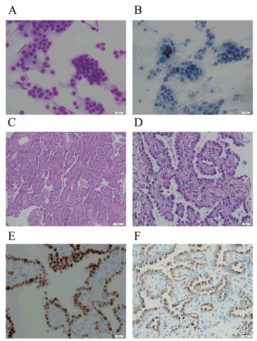
| Figure 1: A case of lung adenocarcinoma had morphologic features of nonmucinous adenocarcinoma in situ (AIS). The Touch preparation slides showed cellular smears with some relative uniform tumor cells with moderate amount of cytoplasm without prominent nucleoli (A, Diff-Quik, 400x; B, Pap stain, 400x). The core tissue demonstrated the neoplastic cells with a bronchial alveolar growth pattern and relative bland nuclei, and there is no evidence of invasion (C, H&E, 100x; D, H&E, 400x). Tumor cells are positive for TTF1 (E, 400x) and Napsin A (F, 400x). |