 |
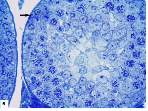
| Figure 5: A photomicrograph of a semithin section in seminiferous tubule of group I showing regular basal membrane and well distinct myoid cell (↑). Normal spermatogenic cells formed of 4-6 layers at different stages of spermatogenesis, Spermatogonia (Sg), primary spermatocytes (Sc) with curled chromatin and spermatids (Sd). Pyramidal Sertoli cells exhibited prominent nucleolus resting on the basement membrane (*). Note the absence of sperms in the non-canalized seminiferous tubule (Toluidine blue, X1000). |