|
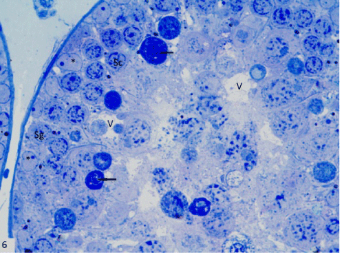
| Figure 6: A photomicrograph of a semithin section in seminiferous tubule of group II revealing intact basement membrane with reduction and disorganization in the spermatogenic layers with complete absence of spermatids. The spermatogonia (Sg), Sertoli (*) and some of the primary spermatocytes (Sc) seemed similar to control. Dense cells with pyknotic nulei were obviously noted in the tubule (↑). Note the presence of vacuoles (v) in between the cells (Toluidine blue, X1000). |