|
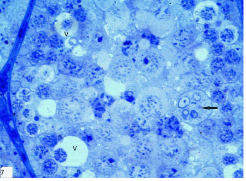
| Figure 7: A photomicrograph of a section in seminiferous tubule of group III revealing vacuoles of different sizes in between and surrounding the spermatogenic cells (v). The spermatogenic cells showed variable nuclear staining affinity. The cells appeared disorganized and dissociated from each other. Note the presence of large cell containing multiple nuclei (↑) and absence of spermatids except for one degenerated spermatid was seen inside this multinucleated cell (Toluidine blue, X1000). |