 |
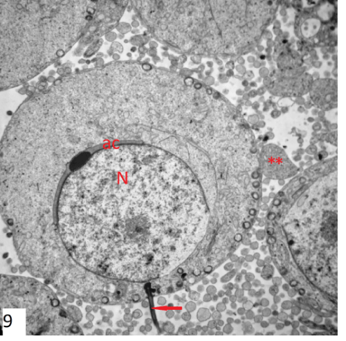
| Figure 9: An electron photomicrograph of a section in the late spermatid of group I showing rounded nucleus (N). The spermiogenesis was manifested by the presence of well developed acrosomal cap and vesicle (ac), flagellum (↑) and many sheded cytoplasmic plebs (**) (X3600). |