 |
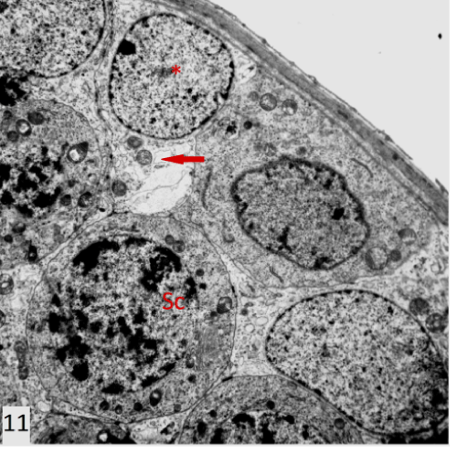
| Figure 11: An electron photomicrograph of a section in the testis of group II showing a primary spermatocyte (Sc) very similar to that of the control. Sertoli cell (*) had indented nucleus with indistict double layer nuclear envelope. The cell had relative electron dense vaculated cytoplasm (↑) (X3600). |