 |
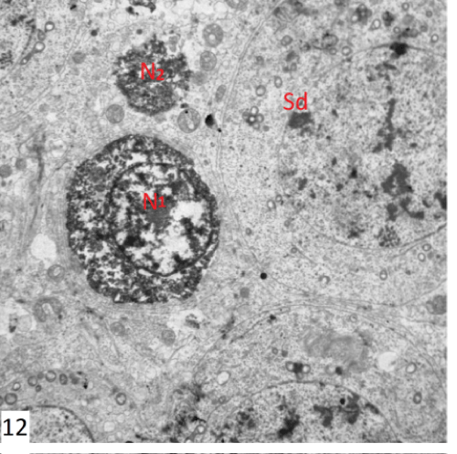
| Figure 12: An electron photomicrograph of a section in the testis of group II showing normal spermatid (Sd). There were two cells which had electron dense nuclei with amalgamation and segmentation of the chromatin clumps (N1 & N2). The cytoplasm of N1 was electron dense and fragmented with loss of the organelles. While the other cell N2 exhibited vaculated cytoplasm with numerous mitochondria (X3600). |