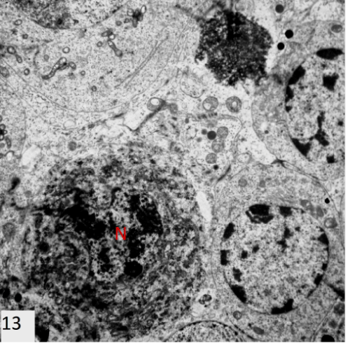
Figure 13:
An electron photomicrograph of a section in the testis of group II showing damaged cell with hyperchromatic irregular nucleus (N) surrounded by degenerated electron dense cytoplasm (X3600).