 |
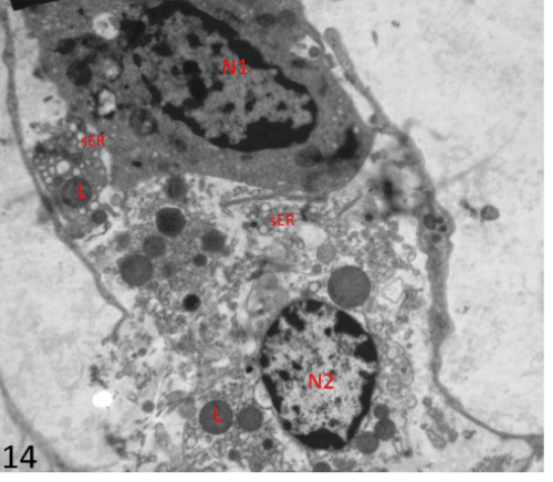
| Figure 14: An electron photomicrograph of a section in the interstitial tissue of the testis of group II showing two Leydig’s cells. One (N1) had irregular oval nucleus and electron dense cytoplasm containing numerous smooth endoplasmic reticulum (sER) and secondary lysosomes (L). There was absence of lipid droplets. N2 was ruptured with dilated smooth endoplasmic reticulum (sER) and lysosomes (L) in the cytoplasm (X4800). |