 |
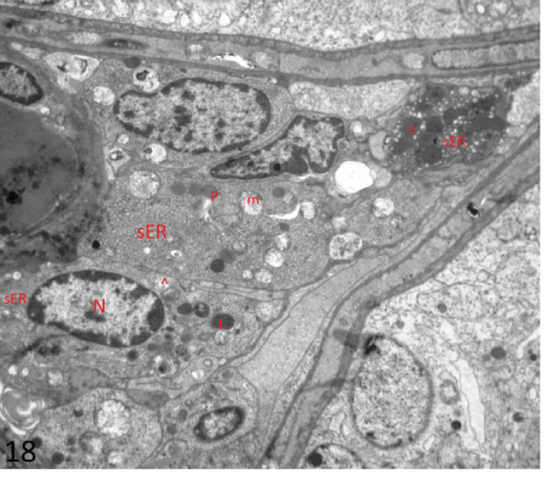
| Figure 18: An electron photomicrograph of a section in the interstitial tissue of testis of group III showing Leydig’s cells. One cell had irregular euchromatic nucleus (N). Its cytoplasm exhibited smooth endoplasmic reticulum (sER), destructed mitochondria (m), few lipid droplets (P) and lysosomes (L). Note the presence of mitochondria with tubular cristae (^) (X4800). |