|
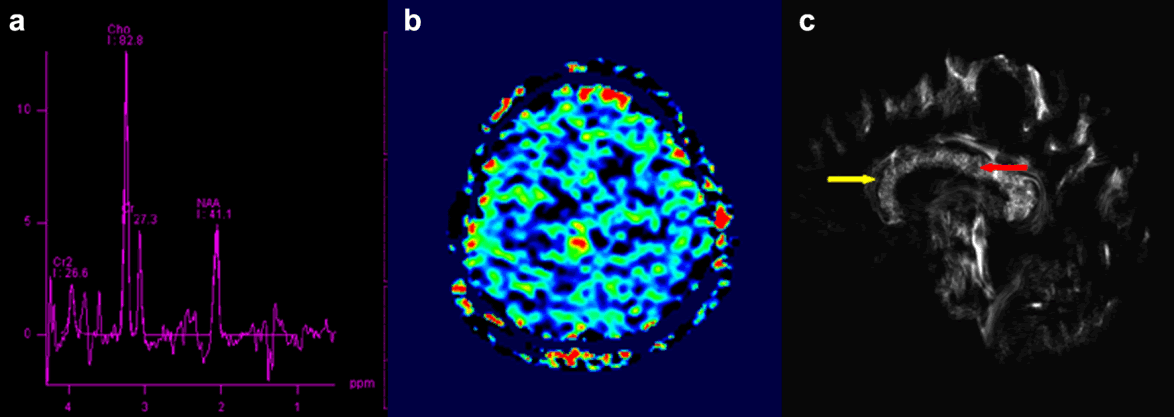
| Figure 2: Multiparametric images from integrated PET/MRI system. (a) Single-voxel MRS shows decreased NAA and elevated choline, which indicates a primary intracranial neoplasm. (b) 2D-ASL indicates the hyperperfusion of the frontal lesion. (c) The track density imaging (TDI) showing the white matter fiber in the body of corpus callosum was disrupted by the tumor infiltration with decreased relative track density (red arrow, 4.4) than the normal region of the genu of corpus callosum (yellow arrow, 10.7). |