 |
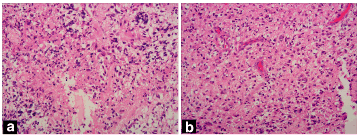
| Figure 3: Histopathological images of the right frontal lesion: (a) Necrosis and microvascular proliferation of glioblastoma. (Stain: hematoxylin-eosin; original magnification: X200). (b) Cellular and diffusely infiltration of tumor cells with rounded hyperchromatic nuclei, perinuclear halos. (Stain: hematoxylin-eosin; original magnification: X200). |