|
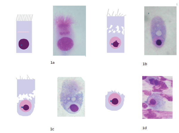
| Figure 1: (a-d): Ciliocytophthoria. We describe the most common morphologic alterations we have observed, assignable to CCP. 1a: Ciliated cell with well-conformed ciliary apparatus, homogenous cytoplasm, and the typical Hypercromatic Supranuclear Stria (HSS). 1b: Ciliated cell with rarefaction of the ciliary apparatus, HSS disappearance, cytoplasmic vacuolization, condensation of the nuclear chromatin with intranuclear halo. 1c: Ciliated cell with coalescence of multiple intracytoplasmic vacuoles. Condensation of the nuclear chromatin with visualization of the nucleolus in the intranuclear halo. 1d: Ciliated cell. “Decapitation” of the apical portion of the ciliated cell, due to the latero-lateral confluence of the cytoplasmic vacuoles. Secondarily to this, it is possible to observe only the caudal portion of the cell, with the nuleus and the nucleolus, all surrounded by a thin cytoplasmic remnant. |