 |
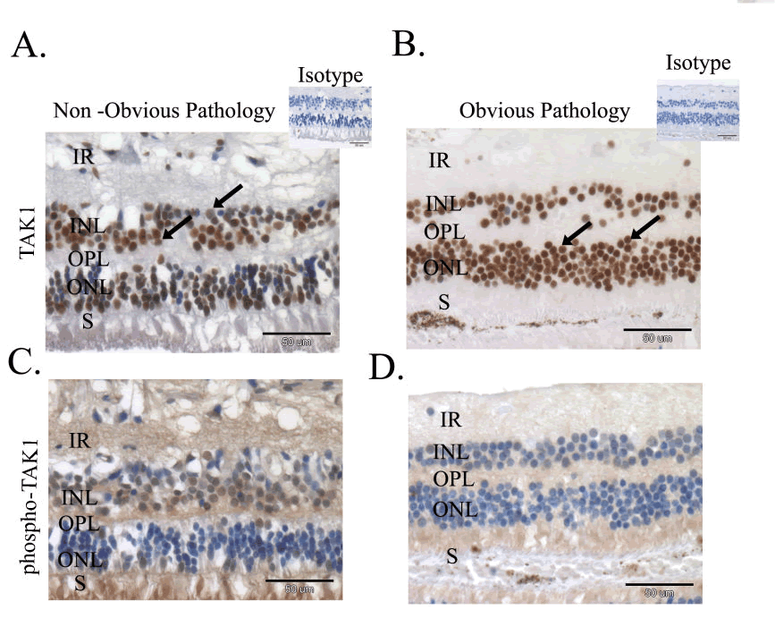
| Figure 1: Aberrant Activity of TAK1 in Human Pathologic Retina. A and B: Retinal specimens, were immunostained with anti-TAK1 antibodies and hematoxylin (TAK1 expression is manifested by brown staining and black arrows), blue- hematoxylin. C and D: Retinal specimens were treated as in A and stained with phospho- Thr 187 TAK antibodies (brown) and hematoxylin (blue), demonstrating aberrant activity of TAK1 in the retina of blind painful eyes. IR-inner retinal layers; INL- inner nuclear layer; OPLouter plexiform layer; ONL- outer nuclear layer; S- photoreceptor segment (Scale bar = 50μm). |