|
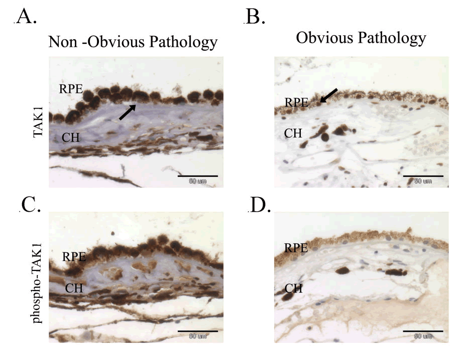
| Figure 2: Attenuated TAK1 Activity in RPE Cells with Pathology. A and B: RPE cells of a retinal specimen were immunostained with TAK1 antibodies and hematoxylin. (TAK1 is manifested by brown staining and black arrows), blue- hematoxylin (N=5). C and D: Retinal specimens treated as in A and stained with phospho-Thr 187 TAK antibodies (brown) and hematoxylin (blue), demonstrating aberrant activity of TAK1 in the RPE cells of blind painful eyes (Scale bar = 50μm). |