 |
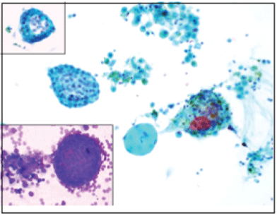
| Figure 1: (Images taken at 200x magnification). Pap-stained smear showing (from L-R) (1) a glandular and tight ball-like structure, with peripheral columnar cells and central stromal-like cells, suggestive of endometrial origin; (2) a collagen ball; (3) clusters of haemosiderin-laden macrophages. Inset pictures show tight balllike clusters of endometrial cells and background red blood cells. |