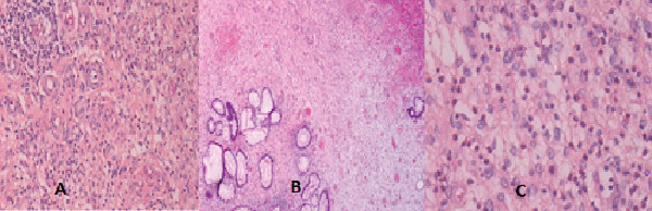
Figure 3:
Characteristic pathology of IFP. Plate A showing broadening of the submucosal layer. Plate B showing prominent capillaries and plate C showing eosinophilic infiltrate.