|
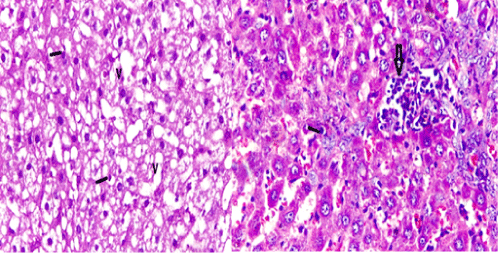
| Figure 3: A photomicrograph of a liver section from adult male rat from Group ΙΙ: (4 wks after treatment of MG), showing degenerated hepatocyte cells with vacuolization (V) with some hepatocytes have small deeply stained nuclei (line) and multifocal areas of minute aggregations of lymphocytes scattered in the hepatic parenchyma (arrow), [H&E X 400]. |