 |
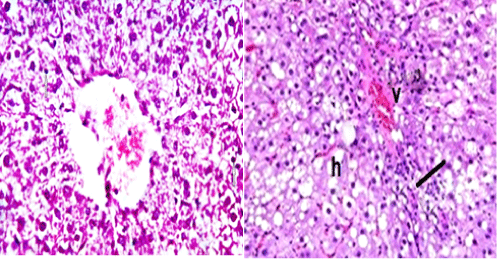
| Figure 5: A photomicrograph of a liver section from adult male rat from Group ΙIΙ: (treated with MG after 2 wks), showing degenerated hepatocyte cells with vacuolization with dark nuclei (arrows) and sinusoidal congestion(v) , degenerating in hepatic cells (h) with infiltration of mononuclear inflammatory cells(line), [H&E X 400]. |