|
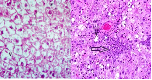
| Figure 6: A photomicrograph of a liver section from adult male rat from Group III: (4 wks after treatment of MG), showing severe degenerated hepatocyte cells with vacuolization (v) with dark nuclei (arrows). Severe inflammatory cells infiltration between hepatocytes (white arrow), [H&E X 400]. |