|
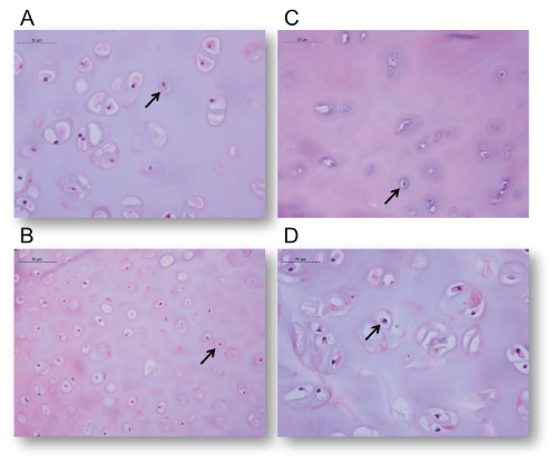
| Figure 3: Optical light microscopy of sculpted cartilage.*High-power, H&E stain showing representative sample of cartilaginous architecture and chondrocyte population in (A) Scalpel, (B) Bovie, (C) Ultrasonic Bone Aspirator and (D) Control groups. Similar density and distribution of viable chondrocytes were noted in all samples. Arrows indicate normal chondrocytes. |