|
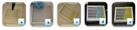
| Figure 4: ELISA screening for Malachite Green and Gentian Violet: a) Addition of standards and samples to the ELISA plate, b) Picture depicting the change in color after the addition of 1X Biotin conjugate to the wells, c) Addition of 1X Streptavidin conjugate to the wells, d) Addition of substrate results in formation of blue color, and e) ELISA plate in its last stage where the addition of stop solution develops yellow color and intensity of this color depicts the presence or absence of the Malachite Green and Gentian Violet. |