|
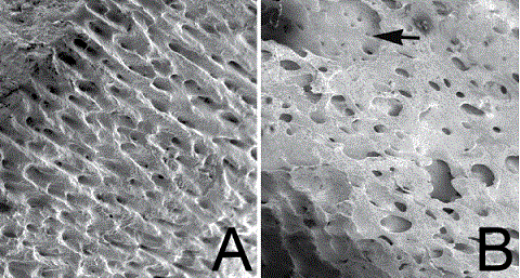
| Figure 3: Scanning electron micrographs of control and trypsintreated samples. Swine bone chips (outer surface of cortical bones; 0.2 g) were collected and examined using SEM: A) Untreated control sample (see Materials and Methods). The control sample showed the outer surface of intact plexiform bone tissue, and B) the trypsin (30 μg/μl) treated sample showed that the exposure of the vascular spaces of plexus (arrow), due to the removal of the surface layer of the bone sample, was observed. Field width: 18 mm. |