|
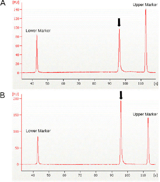
| Figure 4: Results from the Agilent Bioanalyzer 2100 showing electropherograms with the mtDNA Cytb amplicons (arrows). The x-axis on the electropherogram represents the migration time of the amplicon and the y-axis represents the fluorescence intensity of the amplicon. RU: relative fluorescence unit. S: second. Lower marker (15 bp) and upper marker (1500 bp) are the internal size standards. A) Untreated control sample (see Materials and Methods) and B) the trypsin (30 μg/μl) treated sample. |