 |
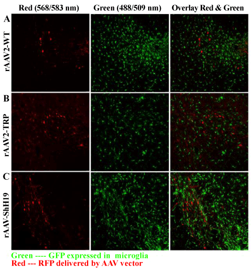
| Figure 4: In vivo transduction of normal brain. Confocal microscopy images of sections of normal brain (no tumor) after AAV vector injections into C57BL/6 CX3CR1/ GFP mice (which expresses GFP in their microglia). Original magnification: 200x. A. rAAV2-WT-CBA-RFP. B. rAAV2-TRP-CBA-RFP. AAV2-TRP is AAV2 capsid with Y(444, 500, 730)F tyrosine mutations (“triple mutant”). C. rAAVShH19- CBA-RFP gene transfer. |