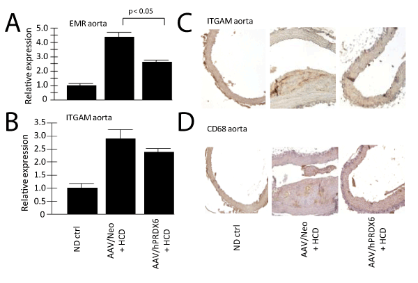
A. shows a QRT-PCR analysis of EMR expression, a marker of macrophages, in the aorta using mRNA from three different animals from each indicated group Note, macrophage levels were significantly lower (p <0.05) in hPRDX6-HCD treated animals than Neo-treated.
B. shows a similar QRT-PCR analysis of ITGAM expression, another macrophage marker. Note, again, macrophage levels trended lower in hPRDX6-HCD treated animals than Neo-treated.
C. shows an immuno-histochemical analysis of ITGAM expression (macrophages). Histologic sections of aorta from the indicated animal groups were analyzed for ITGAM protein using anti-ITGAM antibody. Note that the AAV/hPRDX6-HCD treated animals displayed a much lower brown ITGAM signal than the AAV/Neo-HCD treated animals.
D. shows a similar analysis with anti-CD68 antibody, another marker of macrophages, with similar results as ITGAM.