|
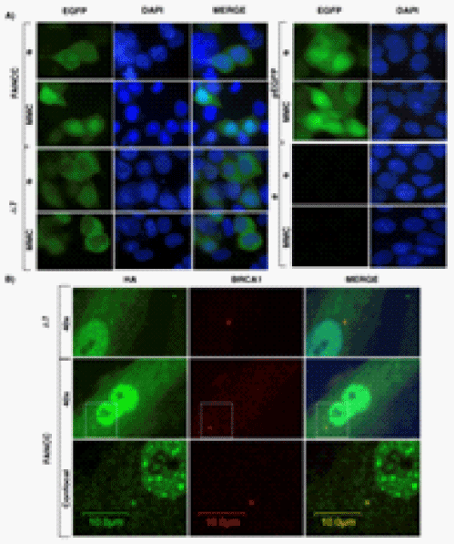
| Figure 5: Cellular localization. (A) Fluorescence microscopy of FANCCEGFP and FANCCΔ7-EGFP transiently transfected in HEK293T cells treated or not with 50 ng/ml of MMC for 16 hours. Counterstaining with DAPI is performed to visualize the nucleus. pEGFP are cells transfected with empty EGFP vector and ø are non transfected cells. (B) Immunofluorescence of FANCC-HA and FANCCΔ7-HA (in green) with BRCA1 (red) in FANCCdeficient cells PD331 infected by retrovirus (using viral vector pMSCVpuro and indicated cDNA). Dashed white squares show the region analyzed by confocal microscopy to confirm co-localization. Scale bars: 100 μm. |