|
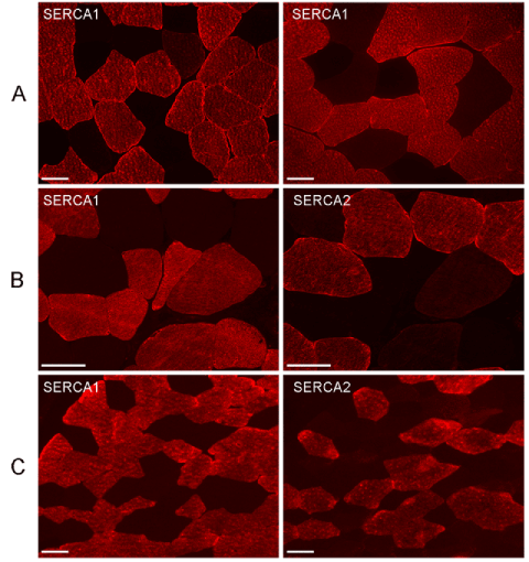
| Figure 4: Immunohistochemistry for SERCA1 and SERCA2. Immunostaining for SERCA1 is similar in muscle of controls (A) and of patients with (B) and without (C) ATP2A1 mutation. Serial sections from muscle biopsy specimen of patients (B, C) show that SERCA1 and SERCA2 antibodies stain type II and type I fibers, respectively. Bars, 50 μm. |