|
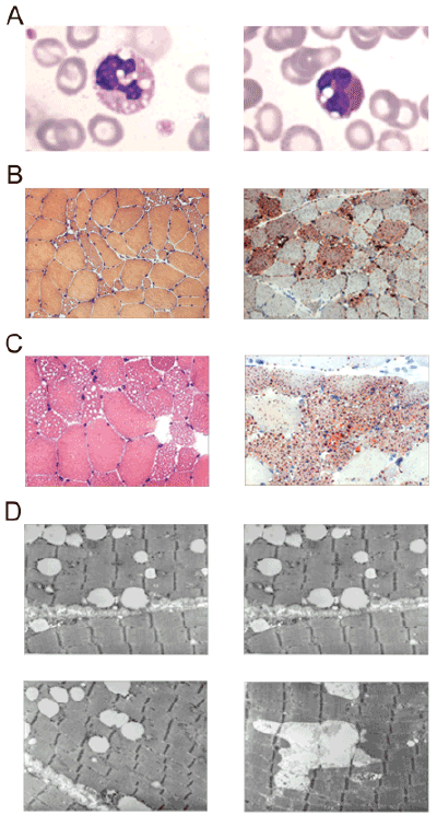
| Figure 2: Histochemical characterization of NLSDM patient. A) Microphotograph of Jordans’ bodies in patient’s buffy coats stained with May- Grünwald Giemsa. B) First muscle biopsy, performed during 2004 and stained with Hematoxylin and Eosin (HE) and ORO, shows vacuolar myopathy with lipid storage; C) Second muscle biopsy, performed during 2005 and stained with HE and ORO, shows vacuolated fibers with a increase of lipid droplets; D) Electron microscopy, performed during 2007, reveals massive line-up of lipid droplets without signs of mitochondrial alteration. |