|
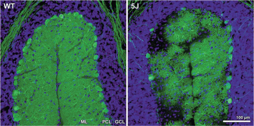
| Figure 3: Purkinje cell degeneration in pcd5J homozygous mice. Here we see cerebellar sections for matched folia from P25 wild-type DBA/2J (WT) and P25 pcd5J homozygous mice (5J) immunostained with an anti-calbindin antibody and counterstained with DAPI. Although WT P25 cerebellum displays numerous Purkinje cell neurons with extensive dendritic arborization visible in the molecular layer (ML), the pcd5J P25 cerebellum reveals pronounced loss of Purkinje cells and dramatic degeneration of surviving Purkinje cells. The degree of Purkinje cell loss and degeneration observed in pcd5J homozygous mice is consistent with the disease’s natural history in pcd5J homozygous mice, suggesting that 5J is a severe allele. ML=molecular layer; PCL=Purkinje cell layer; GCL=granule cell layer. Reproduced with permission from the corresponding author Albert R. La Spada (Chakrabarti et al., 2006). |