|
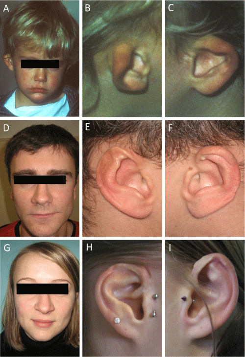
| Figure 1: Photographs of the two affected patients. Frontal view of the patient I.1 at the ages of 1 year (A, B, C) and at adulthood (D, E, F) and of the patient I.2 at adulthood (G, H, I) showing the absence of facial dysmorphia (A, D, G) and the microtia before (B, C) and after surgery (E, F, H, I). |