 |
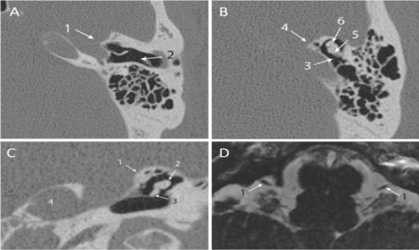
| Figure 3: Radiological images illustrating ear abnormalities of affected patient I.1. (A, B) Axial CT scan showing the bilateral aplasia of the inner ear structures (1), normal external auditory canal (2), hypoplasia of the middle ear (3) and bilateral aplasia of the petrous apex and non-development of the internal auditory canal (4). Incus and Malleus are shown by arrows (5) and (6) respectively. (C) Coronal CT scan of the skull base and petrous bones showing the bilateral absence of the inner ear structures associated with bilateral aplasia of the petrous apex and non-development of the internal auditory canal (1). Incus and Malleus are shown by arrows (2) and (3) respectively. (4) Left jugular foramen. (D) Axial MRI of the cerebellopontine angles in high resolution T2 weighted sequence showing the bilateral absence of the cochleovestibular nerve and the non-development of internal auditory canal. (1) Facial nerves. |