|
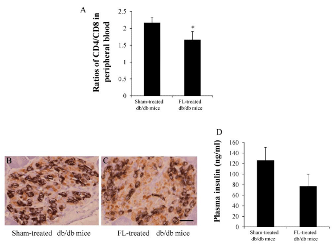
| Figure 4: Ratios of CD4/CD8 and Imaging Analysis in the Pancreata (A) Ratios of CD4/CD8 in the peripheral blood. (B and C) Immunochemistry staining for insulin (brown) and glucagon (black). There was considerably more insulin content in residual beta cells (brown color in C) when compared to sham-treated db/db mice (B). (D) Plasma insulin levels. The results are mean ±SD, n=6 in each group. Scale bar =25μm |