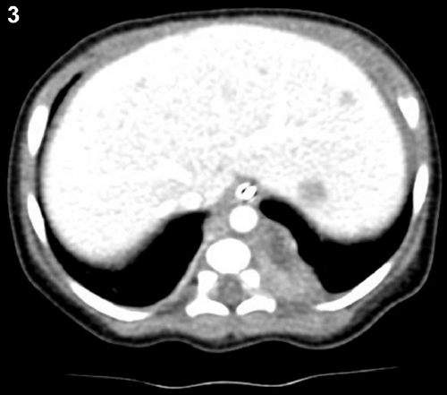
Figure 3:
A section from the abdominal CT conducted during the third week of life. It highlighted calcified liver mass.