|
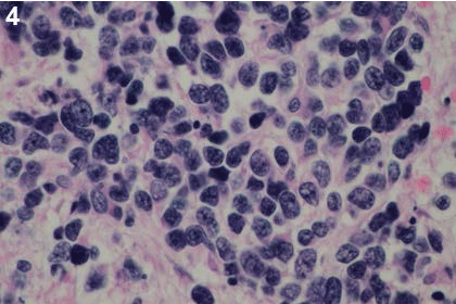
| Figure 4: A microscopic image of the biopsy taken during the second laparotomy after sectioning and staining with Hematoxylin and Eosin. The image facilitates diagnosis of Neuroblastoma (Schwannian stroma poor) poorly differentiated subtype composed of undifferentiated neuroblastic cells with recognizable neuropil (primary magnification x 400). |